


Gonarthrosis, or deforming arthrosis of the knee joint, is a degenerative-dystrophic disease of the hyaline cartilage of the knee, which covers the condyles of the tibia and femur.
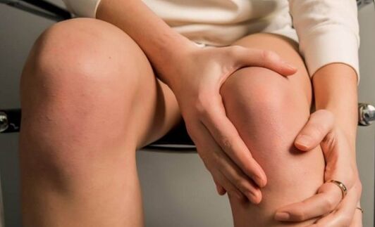
Symptoms of arthrosis of the knee joint develop gradually over the years, and the main manifestation of the disease is stiffness of movements and pain.Among arthrosis of other joints (phalanges of the fingers, shoulder and elbow joints, hip joints), gonarthrosis is the most common disease.
The category of the population most susceptible to arthrosis of the knee joint is women, as well as people over 40 years of age.In some cases, gonarthrosis can develop in athletes (due to increased stress) and young people (against the background of previous injuries).
Causes of gonarthrosis
Osteoarthritis of the knee joint does not develop from a single cause;in most cases, the development of the disease depends on a combination of several negative factors, which worsen with age and can ultimately lead to such a disorder.Medicine identifies the following mechanisms for the onset of osteoarthritis:
Primary – occurs at an older age due to aging of body tissues, as well as against the background of some factors provoking this disease (constant increased stress, heredity, obesity).
Secondary – accounts for 30% of all cases of gonarthrosis, occurring mainly after damage to the meniscus, rupture of ligaments, fracture of the tibia and other injuries.In most cases, symptoms of knee osteoarthritis begin to appear after 3-4 years.However, if the injury is severe enough, it may take 2-3 months for symptoms to appear.
About 7-8% of cases of arthrosis of the knee joint occur when, after forty years, a person begins to actively engage in physical exercise, namely squats and running.But due to age, weakening of the tissue tone of the knee joint and characteristic age-related changes under sudden loads can provoke the appearance of rapid degenerative and dystrophic changes in the joints.
Another reason for the development of gonarthrosis in a person can be concomitant diseases: varicose veins, excess weight, ankylosing spondylitis, gout, psoriatic, rheumatoid or reactive arthritis.
Constant critical loads on the knees, long-term regular climbing of stairs in old age or lifting heavy loads, as well as professional sports can lead to arthrosis of the knee joint.The risk of arthrosis of the knee joint increases significantly in case of genetic weakness of the ligament system, diabetes mellitus and other metabolic disorders, neurological diseases and spinal injuries.
Spasm of the muscles of the front of the thigh is the cause of 50-60% of cases of osteoarthritis of the knee joint.Such muscle spasm may not appear for a long time before knee pain appears, while the person may feel heaviness in the legs, fatigue, and experience lower back pain.When the iliopsoas and rectus femoris muscles are in a constant state of spasm, with age this leads to a "stiffening" of the knees, which limits their freedom of movement.
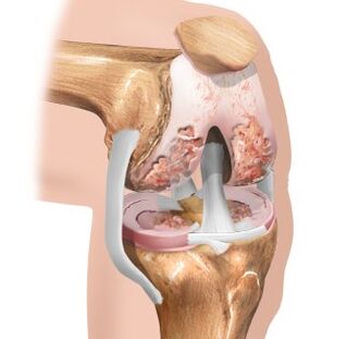
Degrees of gonarthrosis
In most cases, the development of arthrosis of the knee joint occurs in one of the knees.Medicine distinguishes 3 degrees of gonarthrosis depending on the intensity of the pathological processes:
The first is that there is no deformation of the bones of the joint;periodic dull pain in the knee is characteristic after physical stress on the joint.A slight swelling of the joint may develop, which disappears on its own after some time.
The second degree is characterized by an increase in symptoms of arthrosis.The pain is intense and long-lasting and occurs with minimal effort (heavy lifting, walking), and a crunching sound may also appear in the joint.In the second degree of gonarthrosis, a deformation of the knee joint and moderate limitation of movement may develop.
Third degree – presents pronounced symptoms of arthrosis of the knee joint.Significant deformation of the joint and constant pain lead to disturbances in the patient's gait and the mobility of the joint is limited.In the third degree of gonarthrosis, the pain can intensify depending on time and reach such an intensity that it disturbs the patient's sleep, the limitation of mobility is minimized, which makes it impossible to find a comfortable position for the leg.
Diseases and pathologies with which gonarthrosis is confused
Locking of the knee joint in case of damage to the meniscus.
The disease develops after unsuccessful movement, accompanied by a creak of the knee, severe pain appears that disappears after 15 minutes, and the next day swelling of the knee occurs.
Ankylosing spondylitis, gout, joint rheumatism, psoriatic, reactive, rheumatoid arthritis.
To distinguish gonarthrosis from arthritis it is necessary to perform a general blood test.With arthritis, the test results will indicate the presence of an inflammatory process in the body, while with arthrosis there are no changes in the blood test.
Inflammation of the hamstrings.
It develops in most cases in women over 40, when they carry heavy objects or go down stairs.The pain is localized on the inner surface, but the movement of the knee is not limited.
Vascular pain in the knees.
The pain is characterized by symmetry and occurs simultaneously in both knees.Characteristic of young people during a period of accelerated bone growth, they can occur during physical activity, a cold or a change in the weather.To describe their condition, patients use the term “sprained knees.”
Stages of arthrosis of the knee joint
The disease is insidious due to its gradual development.Initially, the patient feels only mild discomfort, pain when moving, going up or down stairs.In some cases, the patient's sensations are reduced to a slight stiffness of the joint and tension in the popliteal region.A feature of arthrosis of the knee joint is considered to be a symptom of initial pain, which consists in the fact that after a sharp transition from a sitting position to movement, a person begins to feel pain during the first steps, which gradually disappears, and then arises again after a heavy load.
The appearance of the knee joint with first degree arthrosis is no different from a healthy knee, only in some cases patients note slight swelling.Also, in some cases, fluid begins to accumulate in the knee joint, it swells and becomes spherical, and synovitis begins to develop, which leads to a feeling of heaviness and limited movement of the joint.
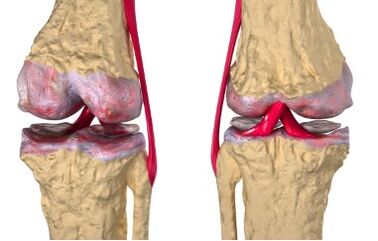
Such changes in the joint in the first degree of arthrosis occur due to disruption of blood circulation in the small intraosseous vessels that nourish the hyaline cartilage.This leads to changes in the surface of the cartilage, dryness, loss of smoothness and the appearance of cracks.For this reason, smooth and soft movement between the cartilages is difficult and is accompanied by constant microtraumas when the cartilages cling to each other.The cartilaginous tissue, being in a constant state of microtrauma, begins to lose its shock-absorbing properties and thin.
The development of pathological processes leads to changes in bone structures.The joint area is flattened, and growths in the form of spines (osteophytes) appear along the edges.The joint capsule degenerates, joint fluid thickens, and the synovial membrane narrows.This leads to a significant deterioration of the nutritional process of the cartilage and accelerates its degeneration.
As the disease progresses to the second stage, the symptoms begin to intensify, the pain occurs even with light loads and is localized along the internal anterior part of the joint.Rest brings relief, but resuming movement causes severe pain.
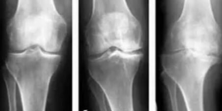
The mobility of the joint is significantly reduced, attempts to bend the leg as much as possible cause sharp pain, and a distinct rough crunch is heard during movement.Synovitis is more common;the joint widens and changes configuration due to a large accumulation of fluid.
The third degree of arthrosis of the knee joint is accompanied by significant deformation of the bones, in which they seem to be pressed into each other.There is practically no cartilaginous tissue and the limitation of joint movement increases.Straightening the joints becomes quite problematic, and the pain begins to bother the patient around the clock, both when walking and at rest.Due to severe deformation of the joints, the legs acquire an O- or X-shape, the gait becomes unstable, sways, and very often the patient cannot do without a cane or crutches.
Diagnosis and treatment of gonarthrosis
During a routine examination, it is impossible to determine whether a patient has first-degree arthrosis of the knee joint.If there is a grade 2 or 3, on examination swelling, edema (with synovitis) is observed, a crunching sound is heard, stiffness of movements is observed, a change in the axis of the limbs is noted, and deformation of the joints is noted.
Today, in addition to radiography, CT and MRI methods are widely used to diagnose arthrosis, thanks to which it is possible to study disorders of bone structures in more detail and examine pathological changes in soft tissues.
Treatment of the disease involves a complex of orthopedic procedures and drug therapy.The orthopedist prescribes mud therapy, therapeutic exercises, massages and physiotherapy.
Pharmacological treatment involves the use of chondroprotectors, these are drugs that replace synovial fluid.In some cases, intra-articular injection of steroid hormones is performed.Patients are also recommended to undergo sanatorium-resort treatment.
If treatment of a young patient with severe limitation of movement and pain is ineffective, a joint replacement method can be used, which involves a rehabilitation period of 3-6 months.
What happens to the knee joint with osteoarthritis?
Articular cartilage plays an important role in human movement and is a smooth, elastic and durable covering, thanks to which the jointed bones move smoothly relative to each other and also absorb and distribute the load when walking.
But with excessive regular loads, genetic predisposition, metabolic disorders in the body, prolonged muscle spasms and injuries, the cartilage loses its smoothness and begins to thin.The soft sliding of the jointed bones is replaced by strong friction and the first degree of gonarthrosis develops, in which the cartilage loses its shock-absorbing properties.
The degradation process continues to progress and poor shock absorption leads to flattening of bone surfaces with the formation of osteophytes in the form of bone growths.In this case the disease already has a second degree and is accompanied by degeneration of the synovial membrane and joint capsule.The lack of pumping and movement atrophies the structure of the knee joint, the consistency of the knee fluid becomes more viscous, the process of cartilage nutrition is disrupted, which leads to an even greater worsening of the patient's condition.
The thinning of the cartilage leads to a reduction in the distance between the articulating bones by up to 80%.While walking, due to abnormal friction and the absence of shock absorbers, destructive processes in the joint increase rapidly, this quickly leads to the development of the third degree of arthrosis with pronounced symptoms:
Pain during movements, especially when going up or down stairs.
Pain both during physical activity and at rest, morning stiffness.
The patient begins to limp, trying to spare the painful joint.
Severe cases of the disease require the use of crutches or a cane.
The third degree of arthrosis of the knee joint is characterized by an almost complete absence of cartilaginous tissue, which leads to a minimal decrease in joint mobility.Therefore, no magical methods, supermedicines or ointments can restore worn cartilage tissue, and given the degree of bone deformation, normal functioning of the joint is impossible.In this case only surgery can help.
Gymnastics for arthrosis of the knee joint
Any treatment option for arthrosis of the knee joint should be carried out only on prescription.Therapeutic gymnastics involves slow, measured exercises that exclude squats, twisting the joint and jumping.It is preferable to do gymnastics in the morning, sitting or lying down, for 20 minutes, repeating each exercise 10 times.
The main goals of therapeutic exercises are to relax muscle spasms that cause pain, increase blood flow to the joint, slow the progression of the disease, and prevent further destruction of the cartilage.During the period of exacerbation of the disease, physical exercise is prohibited.

Lying on your back, you can perform the bicycle exercise, but you need to straighten your legs parallel to the floor, make circular movements with your feet, move your legs to the side, sliding them alternately along the floor, stretch your feet for a count of 10.
Sitting on a chair with your legs lowered, straighten your legs, simultaneously bending your feet and hold this position, counting to 10, alternately pull each knee to your stomach with your hands and slowly return to the starting position.
Concentrating on the wall, standing on the floor, alternately swing your legs back and forth.
Placing the straightened leg on a chair, perform inclined movements of an elastic nature, resting your hands on the thigh, as if trying to straighten the leg more.
Lying on your stomach, alternately lift your straight leg and hold it for a count of 3.
Sitting on the floor, spread your legs to the sides, moving them along the floor, bring your knees closer to your stomach as you inhale, and return them to the starting position as you exhale.